This content is only available in English
Core Facility Cell Analysis
The Core Facility Cell Analysis (CFCA) is a cytometry and imaging facility that provides a convenient and guided access to advanced cell analysis technologies and fosters the benefits of these resources to researchers in and around TranslaTUM. The facility hosts a high-end flow cytometer and cell sorter, a high throughput imaging cytometer, an advanced confocal microscope as well as a mass cytometer for the exploratory edge. This collection of resources along with the available technical expertise serves as the ultimate foundation for rapid and efficient in-depth phenotypic and functional analysis at cellular dimension. Strategically located on the second floor in TranslaTUM, CFCA not only conducts biologically relevant projects in fundamental, pre-clinical and translational research but also contributes to in-house technology development and engineering by offering commercial state of art technologies as reference. CFCA users benefit from project designing guidance, service-based or (post-training) self-usage of instruments as well as data analysis assistance.
Instruments
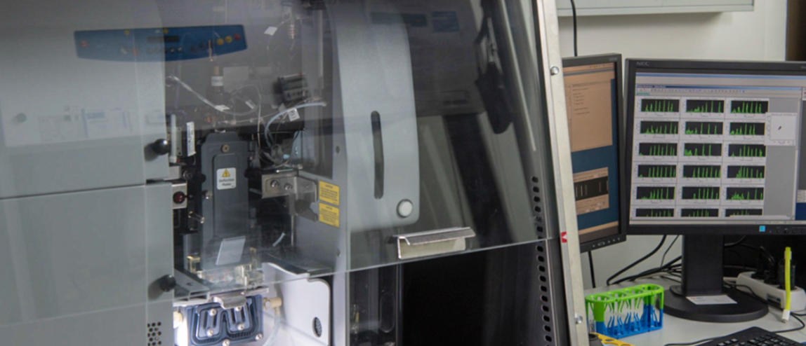
State-of-the-art 5-laser / 20 parameter flow cytometry cell sorter placed in a biosafety cabinet enables service-based sterile cell sorting. Samples up to Bio II/S2 can be processed here, and sterile cell culture facilities exist in the same room for sensitive samples.
- Sorting from 5 ml or 15 ml sample tubes
- Up to 4-way sorting (in 1.5 ml and 5 ml tubes), up to 2-way sorting (in 15 ml and 50 ml tubes) or 1-way sorting (plus index sorting for single cell sorts) into plates
- Temperature of sample tube holder and collection tube holder adjustable from 4°C to 37°C
This instrument is located in room 22.2.45 (Bio II/S2)
For detailed optical configuration, click here


A high-end 4-laser / 17-parameter flow cytometry cell sorter enables service-based sterile cell sorting. The specialized fluidics and optics maximize signal detection while minimizing cross-talk, ensuring high-quality results. Samples up to Bio I/S1 can be processed here.
- Sorting from 5ml or 15ml sample tubes
- Up to 4-way sorting (in 1.5ml and 5ml tubes), up to 2-way sorting (in 15ml and 50ml tubes) or 1-way sorting (plus index sorting for single cell sorts) into plates
- Adjustable temperature for both sample tube holder and collection tube holder, ranging from 4°C to 37°C
This instrument is located in room 22.2.44 (Bio I/S1)
For detailed optical configuration, click here
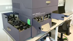
An advanced cell analyzer equipped with 5 lasers to support complex multicolor flow cytometry assays. Up to 18 fluorescent parameters can be simultaneously and rapidly analyzed here. Due to the specialized fluidics and optics of this instrument, users benefit from maximized signal detection accompanied by minimum cross talk. A separate analysis system with FlowJo software is offered for users of the core facility and can be reserved via the online booking system.
This instrument is located in room 22.2.44 (BioII/S1)
For detailed optical configuration click here
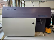
A cell analyzer equipped with 3 lasers to support multicolor flow cytometry assays. Up to 8 fluorescent parameters can be simultaneously and rapidly analyzed here. Due to this instrument's specialized fluidics and optics, users benefit from maximized signal detection accompanied by minimum cross-talk. The core facility is equipped with two BD FACSCanto II instruments. A separate analysis system with FlowJo software is offered for core facility users and can be reserved via the online booking system.
This instrument is located in room 22.2.44 (BioII/S1)
For detailed optical configuration click here
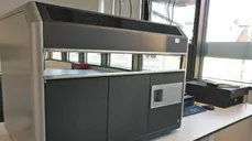
Amnis Image Stream X Mark II Imaging Flow Cytometer combines the best from the worlds of flow cytometry and microscopy. It is capable of fast and sensitive phenotypic analysis together with crisp imagery of every cell directly in the fast flow, permitting scientists to gather high throughput cellular localization data of biomolecules of interest. The data generated from the imaging flow cytometer can be analyzed with Ideas analysis software available on a separate analysis system.
This instrument is located in room 22.2.44 (BioII/S1)
For detailed optical configuration click here
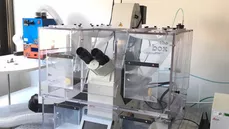
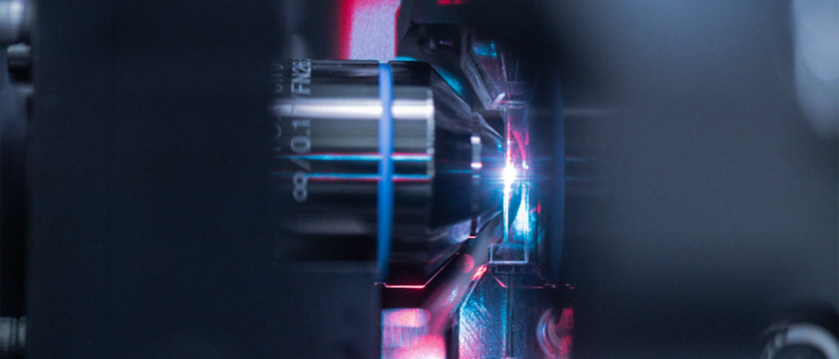
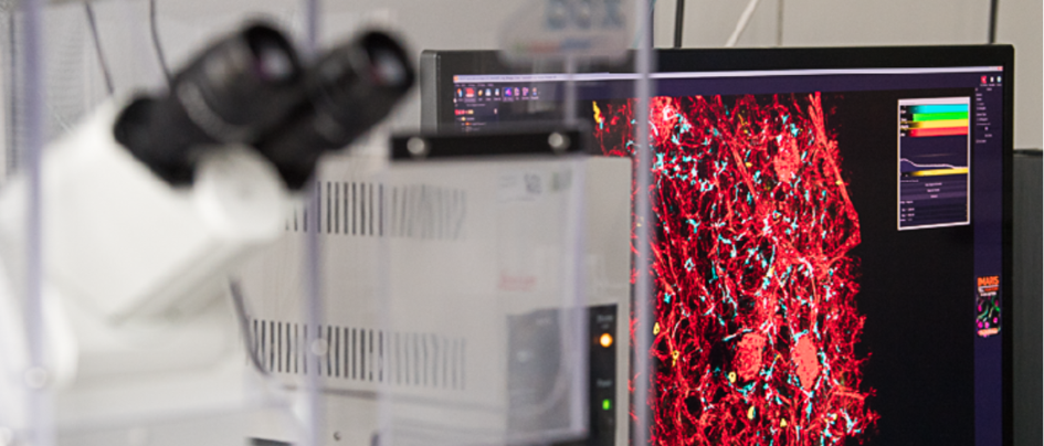
High-resolution multi-color imaging of biological processes is feasible on the Leica SP8 Confocal microscope. The system is equipped with a white light (470-670 nm) and a 405 diode laser, motorized stage, incubator system for full climate control during live cell measurements and deconvolution. Sample like cell monolayers, tissue sections, organoids and more can be imaged post-fixation or living with the confocal. The 3D and 4D data produced can be visualized and analyzed using the Imaris software available on a separate analysis system.
This instrument is located in room 22.2.44 (BioII/S1)
For detailed optical configuration click here
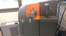
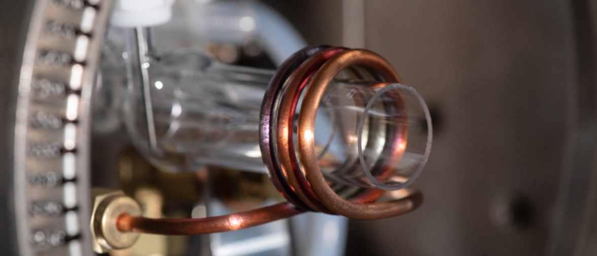
The mass cytometry based analysis platform is offered solely on collaboration basis. Switching from a fluorescence based to a mass spectrometry based detection method enables the parallel detection of 40+ different cell parameters. By labeling cells with metal-tagged antibodies that are measured by plasma time-of-flight technology (TOF), this technique dramatically increases the signal quality and facilitates the collection of significantly more data per cell whereby reducing the need of large amounts of sample material.
This instrument is located in room 22.2.50 (BioII/S1)
Access and Booking System
New project or first cell sorting service
- Contact us and fix a meeting to share the project related details and communicate your experimental/sort requirements.
- Get recommendation on which instruments would be the most suitable for the project and also plan a schedule.
- All questions related to experimental/panel design, sort time needed, sample concentration, nozzle to use, etc. will be talked over.
- If you don’t have a user account for the Core Facility Cell Analysis booking system, the core facility contact will assist.
Regular Users
- Reserve your booking appointment in the Core Facility Cell Analysis online booking system.
- For a sort appointment, fill out the new CFCA sort request form and email to Marie Goess at least 1 day before the scheduled sort. In case of S2 sorts, the signed z-form should also be emailed.
Trainings
- Trainings are offered on a regular basis for independent self-usage of analyzers and microscope.
- Email or call us to include you on the waiting list and we will get back to you once enough aspirant trainees have enrolled.
- The training session is followed by a practice and assessment session. Access to the online booking system and the instrumental access will be granted after the assessment session is successfully passed.
- Flow cytometry theory, FlowJo basics and compensation trainings are also provided and spot on the next session can be requested for.
Appointments can be conveniently reserved via the Core Facility Cell Analysis online booking system after you have passed a self-usage instrument training or booked a service. Access to resources is granted based on the training you have received. If you get trained for a new instrument or have any other personal detail changes (affiliation, designation, contacts, etc.) then please inform the core facility staff immediately for updating your details.
Staff
